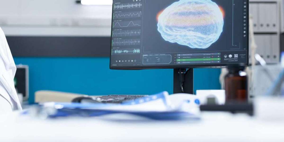
Brain mapping assists researchers to know how the brain works. It shows which parts of the brain do what. Also, it helps doctors find problems in the brain. Today, there are many brain mapping techniques. Every technique functions not in the same way. Let’s discuss the most familiar techniques.
1. MRI (Magnetic Resonance Imaging):
Firstly, MRI is one of the most common using brain mapping Las Vegas equipment. It uses strong magnets and radio waves. As a result, it creates clear pictures of the brain. Also, it does not use harmful radiation. Moreover, MRI shows both the structure and size of the brain. Doctors use it to find brain tumors, strokes, and injuries. In short, MRI is safe, clear, and widely used.
2. fMRI (Functional MRI):
Secondly, fMRI is similar to MRI, but it does more. It shows brain activity. How? It tracks blood flow in the brain. When one part of the brain works, more blood goes there. Because of this, fMRI helps doctors see which brain part is active during a task. For instance, it may display what takes place in the brain while somebody talks or thinks. So, fMRI is very supportive for scheduling brain surgery or examining brain function.
3. CT scan (Computed Tomography):
Another technique is the CT scan. It utilizes X-rays to produce photographs of the brain. Afterward, a computer merges these pictures to produce a 3D vision. CT scans are very speedy and useful in emergencies. For example, they can quickly find bleeding or injury in the brain. However, they use more radiation than MRI. So, doctors use them only when needed. Yet, they are very valuable in most instances.
4. PET Scan (Positron Emission Tomography):
Next, we have the PET scan. It shows how the brain uses energy. The patient is given a small amount of radioactive sugar. Then, the scanner checks how the brain uses it. As functioning brain parts consume extra sugar, PET scans indicate which parts function properly. This aids in analyzing ailments like Alzheimer’s or epilepsy. Furthermore, PET scans may see brain tumors. However, PET scans are costly and use radiation. Even so, they are powerful tools for deep brain study.
5. EEG (Electroencephalography):
EEG is another popular method. It records the brain’s electrical signals. Tiny wires called electrodes are placed on the head. Then, the machine picks up brain waves. Also, EEG is quick, easy, and safe. It is utilized frequently to check for seizures and sleep disorders. For instance, in epilepsy, EEG indicates unusual signs. Although EEG does not display pictures, it informs us how the brain is functioning in real-time. So, it is very beneficial.
6. MEG (Magnetoencephalography):
Likewise, MEG is similar to EEG. But it measures magnetic fields instead of electric signals. The brain gives off tiny magnetic fields when it works. MEG can find active brain areas quickly and clearly. This makes it great for brain research and surgery planning. Though, it is very costly and not broadly accessible.
7. NIRS (Near-Infrared Spectroscopy):
Now let’s look at NIRS. It is a newer method. It uses light to study brain activity. A special device shines near-infrared light into the head. Various light is absorbed, and several is revealed in return. This displays how widely oxygen the brain is consumed. As an outcome, we may look at which parts of the brain are functioning. NIRS is harmless and simple to utilize. Also, it is helpful for babies and people who cannot stay still. Though it is not as clear as MRI or fMRI, it is still useful.
8. Brain Stimulation Techniques:
Some techniques do more than just measure. They also affect brain activity. For example, TMS (Transcranial Magnetic Stimulation) uses magnets to change brain signals.
TMS can help find brain functions. Also, it is used to treat depression. Another method is TDCS (Transcranial Direct Current Stimulation). It uses a small electric current on the scalp.
These techniques are both used in research and therapy. They help us learn about the brain and even improve it.
9. Brain Mapping with Surgery:
In some cases, brain mapping is done during surgery. This is called intraoperative brain mapping. It helps doctors avoid important brain areas while removing a tumor. The patient may be awake. This way, doctors can test speech, movement, or memory. As a result, the surgery is safer. Also, the patient can keep important abilities.
10. Genetic and Molecular Brain Mapping:
Lastly, scientists also map the brain at the genetic level. They study how genes affect brain structure and function. They also use chemical tracers to find brain connections. Though this is complex, it gives deep insights. It can help us understand brain diseases and find new treatments.
Conclusion:
In conclusion, brain mapping Las Vegas is a key part of brain science. It uses many different tools. Each one has its own benefits. MRI and fMRI give clear pictures. CT scans are fast. PET shows energy use. EEG and MEG show brain signals. NIRS uses light. TMS and TDCS can even change brain activity. Also, brain mapping during surgery helps protect the brain. And genetic mapping looks even deeper. Therefore, these tools are very important. Above all, brain mapping brings us closer to understanding the most complex part of our body, the brain.